The Endoplasmic Reticulum
22.01

There are two regions of the ER that differ in both structure and function. One region is called rough ER because it has ribosomes attached to the cytoplasmic side of the membrane. The other region is called smooth ER because it lacks attached ribosomes. Typically, the smooth ER is a tubule network and the rough ER is a series of flattened sacs.
The rough ER manufactures membranes and secretory proteins. In leukocytes (leuk-) the rough ER produces antibodies (anti-). In pancreatic cells the rough ER produces insulin. The rough and smooth ER are usually interconnected and the proteins and membranes made by the rough ER move into the smooth ER to be transferred to other locations.
 Rough Endoplasmic Reticulum
The smooth ER has a wide range of functions including carbohydrate and lipid synthesis. It serves as a transitional area for vesicles that transport ER products to various destinations. In liver cells the smooth ER produces enzymes that help to detoxify certain compounds. In muscles the smooth ER assists in the contraction of muscle cells and in brain cells it synthesizes male and female hormones.

ribosome
21.23
 Ribosomes are organelles that consist of RNA an proteins. They are responsible for assembling the proteins of the cell. Depending on the protein production level of a particular cell, ribosomes may number in the millions.
Distinguishing Characteristics:
Ribosomes are typically composed of two subunits: a large subunit and a small subunit. Ribosomal subunits are synthesized by the nucleolus. These two units join together when the ribosome attaches to messenger RNA to produce a protein in the cytoplasm.Location in the Cell:
There are two places that ribosomes usually exist in the cell: suspended in the cytosol and bound to the endoplasmic reticulum. These ribosomes are called free ribosomes and bound ribosomes respectively. In both cases, the ribosomes usually form aggregates called polysomes.Free ribosomes usually make proteins that will function in the cytosol, while bound ribosomes usually make proteins that are exported or included in the cell's membranes. Interestingly enough, free ribosomes and bound ribosomes are interchangeable and the cell can change their numbers according to metabolic needs.
 Ribosome structure indicating small subunit (A) and large subunit (B). Side and front view.
1 Head
2 Platform
3 Base
4 Ridge
5 Central protuberance
6 Back
7 Stalk
A tiny organelle that is the site of protein synthesis (protein translation) in the living cell. Ribosomes are complex, bead-like structures composed of about 40% protein and 60% ribosomal RNA (rRNA). In eukaryotes, ribosomes are made of four strands of RNA and are often attached to the membranes of the endoplasmic reticulum to form rough ER. In prokaryotes, they are made of three strands of RNA and occur free in the cytoplasm.
Eukaryote ribosomes are produced and assembled in the nucleolus. Three of the four strands are produced there, but one is produced outside the nucleolus and transported inside to complete the ribosome assembly. Ribosomal proteins enter the nucleolus and combine with the four strands to create the two subunits that will make up the completed ribosome. The ribosome units leave the nucleus through the nuclear pores and unite once in the cytoplasm. Some ribosomes will remain free-floating in the cytoplasm, creating proteins for the cell's use. Others will attach to the endoplasmic reticulum and produce the proteins that will be "exported" from the cell.
Protein synthesis requires the assistance of two other RNA molecules. Messenger RNA (mRNA) provides instructions from the cellular DNA for building a specific protein. Transfer RNA (tRNA) brings the protein building blocks, amino acids, to the ribosome. Once the protein backbone amino acids are polymerized, the ribosome releases the protein and it is transported to the Golgi apparatus. There, the proteins are completed and released inside or outside the cell. For more details of the role played by ribosomes in protein synthesis, see protein translation.
Mitochondria
21.11
Mitochondria are the cell's power producers. They convert energy into forms that are usable by the cell. They are the sites of cellular respiration which ultimately generates fuel for the cell's activities.

What are their distinguishing characteristics?
Mitochondria are bounded by a double membrane. Each of these membranes is a phospholipid bilayer with embedded proteins. The outermost membrane is smooth while the inner membrane has many folds. These folds are called cristae. The folds enhance the "productivity" of cellular respiration by increasing the available surface area.

Muscle Cell Mitochondria, Copyright Dennis Kunkel.
The double membranes divide the mitochondrion into two distinct parts: the intermembrane space and the mitochondrial matrix. The intermembrane space is the narrow part between the two membranes while the mitochondrial matrix is the part enclosed by the innermost membrane. Several of the steps in cellular respiration occur in the matrix due to its high concentration of enzymes.

Mitochondrion with matrix, Image courtesy of The Virtual Cell.
Mitochondria are semiautonomous (semi- auto-) in that they can divide and grow to make more of themselves. They also have their own DNA and ribosomes.
Share your opinions
The Cell-Cell Structure
20.53
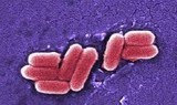
Eukaryotic Cells and Prokaryotic Cells
There are two primary types of cells: eukaryotic cells and prokaryotic cells. Eukaryotic cells are called so because they have a true nucleus. The nucleus, which houses DNA, is contained within a membrane and separated from other cellular structures. Prokaryotic cells however have no true nucleus. DNA in a prokaryotic cell is not separated from the rest of the cell but coiled up in a region called the nucleoid.As organized in the Three Domain System, prokaryotes include archaeans and bacteria. Eukaryotes include animals, plants, fungi and protists. Typically, eukaryoitc cells are more complex and much larger than prokaryotic cells. On average, prokaryotic cells are about 10 times smaller in diameter than eukaryotic cells.
Eukaryotes grow and reproduce through a process called mitosis. In organisms that also reproduce sexually, the reproductive cells are produced by a type of cell division called meiosis. Most prokaryotes reproduce through a process called binary fission. During binary fission, the single DNA molecule replicates and the original cell is divided into two identical daughter cells.
Both eukaryotic and prokaryotic organisms get the energy they need to grow and maintain normal cellular function through cellular respiration. Cellular respiration has three main stages: glycolysis, the citric acid cycle, and electron transport. In eukaryotes, most cellular respiration reactions take place within the mitochondria. In prokaryotes, they occur in the cytoplasm and/or within the cell membrane.
The Cell-Cell Structure
There are also many distinctions between eukaryotic and prokaryotic cell structure. The following table compares the cell structures found in a typical prokaryotic cell to those found in a typical animal eukaryotic cell.Cell Structure Comparison
| Eukaryotic and Prokaryotic Cell Structure | |||
| Cell Structure | Prokaryotic Cell | Typical Animal Eukaryotic Cell | |
| Cell Wall | Yes | No | |
| Centrioles | No | Yes | |
| Chromosomes | One long DNA strand | Many | |
| Cilia or Flagella | Yes, simple | Yes, complex | |
| Endoplasmic Reticulum | No | Yes (some exceptions) | |
| Golgi Complex | No | Yes | |
| Lysosomes | No | Common | |
| Mitochondria | No | Yes | |
| Nucleus | No | Yes | |
| Peroxisomes | No | Common | |
| Cell Membrane | Yes | Yes | |
| Ribosomes | Yes | Yes | |