Tampilkan postingan dengan label anatomy. Tampilkan semua postingan
Tampilkan postingan dengan label anatomy. Tampilkan semua postingan
Anatomy Of Frog
04.43
Frog oval cavity

a. Esophagus opening
b. Vomerine teeth
1. Grove of the maxilla
2. Maxillary teeth
3. Internal nare
4. Eye ball
5. Eustachian tube opening
6. Jaw ridge
7. Underside of the tongue
8. Glottis
9. Cut muscle and bone
Female Frog Internal Anatomy
 1. Liver lobes
2. Heart
3. Ovary with eggs
4. Gall bladder
5. Small intestine
6. Stomach
7. Oviduct
Male Frog Internal Anatomy

a. Large intestine
b. Small intestine
1. Lung lobes
2. Heart
3. Liver lobes
4. Gall bladder
5. Stomach
6. Small intestine
7. Testis
8. Fat body
9. Urinary bladder
Anatomy of the Heart: Aorta
17.56
What is the aorta?
The aorta is the largest artery in the body. Arteries are vessels that carry blood away from the heart. The aorta arises from the left ventricle of the heart, forms an arch, then extends down to the abdomen, where it branches off into two smaller arteries.Function of the Aorta
The aorta carries and distributes oxygen rich blood to all arteries. Most major arteries branch off from the aorta, with the exception of the main pulmonary artery.Structure of the Aortic Walls
The walls of the aorta consist of three layers. They are the tunica adventitia, the tunica media, and the tunica intima.These layers are composed of connective tissue, as well as elastic fibers. These fibers allow the aorta to stretch to prevent over-expansion due to the pressure that is exerted on the walls by blood flow.
Branches of the Aorta
- Ascending Aorta - Extends from the left ventricle of the heart.
- Coronary Arteries
- Aortic Arch
- Brachiocephalic Artery
- Right Common Carotid Artery
- Right Subclavian Artery
Left Common Carotid Artery
Left Subclavian Artery - Right Common Carotid Artery
- Descending Aorta
Chest Region:
- Visceral Branches - Supply blood to the lungs, pericardium, lymph nodes, and esophagus.
- Parietal Branches - Supply blood to the chest muscles, diaphragm, and spinal cord.
Abdominal Region:
- Celiac Artery
- Left Gastric Artery - Supplies blood to the esophagus and portions of the stomach.
- Hepatic Artery - Supplies blood to the liver.
- Splenic Artery - Supplies blood to the stomach, spleen, and pancreas.
- Renal Arteries - Supplies blood to the kidneys.
- Ovarian Arteries - Supplies blood to the female reproductive organs.
- Testicular Arteries - Supplies blood to the male reproductive organs.
- Femoral Arteries - Supply blood to the lower legs and feet.
Superior Mesenteric Artery - Supplies blood to the intestines.
Inferior Mesenteric Artery - Supplies blood to the colon.
Common Iliac Artery - Visceral Branches - Supply blood to the lungs, pericardium, lymph nodes, and esophagus.
Capillary
08.55
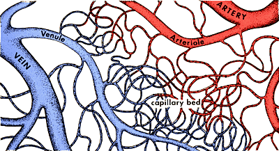
What Is a Capillary?
A capillary is an extremely small blood vessel located within the tissues of the body, that transports blood from arteries to veins. Capillaries are most abundant in tissues and organs that are metabolically active. For example, muscle tissues and the kidneys have a greater amount of capillary networks than do connective tissues.
Capillary Size
Capillaries are so small that red blood cells can only travel through them in single file. Capillaries measure in size from about 5-10 microns in diameter. Capillary walls are thin and are composed of endothelium (a type of simple squamous epithelial tissue). Oxygen, carbon dioxide, nutrients and wastes are exchanged through the thin walls of the capillaries.
Capillary Microcirculation
Capillaries play an important role in microcirculation. Microcirculation deals with the circulation of blood from the heart to arteries, to smaller arterioles, to capillaries, to venules, to veins and back to the heart.
The flow of blood in the capillaries is controlled by structures called precapillary sphincters. These structures are located between arterioles and capillaries and contain muscle fibers that allow them to contract. When the sphincters are open, blood flows freely to the capillary beds of body tissue. When the sphincters are closed, blood is not allowed to flow through the capillary beds. Fluid exchange between the capillaries and the body tissues takes place at the capillary bed.
The flow of blood in the capillaries is controlled by structures called precapillary sphincters. These structures are located between arterioles and capillaries and contain muscle fibers that allow them to contract. When the sphincters are open, blood flows freely to the capillary beds of body tissue. When the sphincters are closed, blood is not allowed to flow through the capillary beds. Fluid exchange between the capillaries and the body tissues takes place at the capillary bed.
Ventricles of the Heart
08.53

Ventricles of the Heart
The heart is divided into four chambers that are connected by heart valves. The lower two chambers of the heart are called ventricles. They are separated by a septum into the left ventricle and the right ventricle. The upper two heart chambers are called atria. Atria receive blood returning to the heart from the body and ventricles pump blood from the heart to the body. Due to the force needed to pump blood to the body, ventricles have thicker walls than do atria.
Function of the Ventricles
The ventricles of the heart function to pump blood to the entire body.
- Right ventricle: Receives blood from the right atrium and pumps it to the main pulmonary artery. The main pulmonary artery extends from the right ventricle and branches into left and right pulmonary arteries, which extend to the lungs. Here oxygen-poor blood picks up oxygen and is returned to the heart via the pulmonary veins.
- Left ventricle: Receives blood from the left atrium and pumps it to the aorta. The aorta carries and distributes oxygen-rich blood to the rest of the body.
Vein
08.47

A vein is an elastic blood vessel that transports blood from various regions of the body to the heart. Veins can be categorized into four main types: pulmonary, systemic, superficial, and deep veins.
Pulmonary veins carry oxygenated blood from the lungs to the left atrium of the heart. Systemic veins return deoxygenated blood from the rest of the body to the right atrium of the heart. Superficial veins are located close to the surface of the skin and are not located near a corresponding artery. Deep veins are located deep within muscle tissue and are typically located near a corresponding artery with the same name (for example coronary arteries and veins).
A vein can range in size from 1 millimeter to 1-1.5 centimeters in diameter. The smallest veins in the body are called venules. They receive blood from the arteries via the arterioles and capillaries. The venules branch into larger veins which eventually carry the blood to the largest veins in the body, the vena cava. The blood is then transported from the superior vena cava and inferior vena cava to the right atrium of the heart.
Artery
08.34
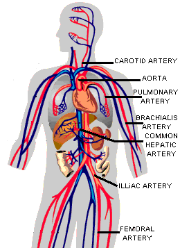 An artery is an elastic blood vessel that transports blood away from the heart. There are two main types of arteries: pulmonary arteries and systemic arteries.
Pulmonary arteries carry blood from the heart to the lungs where the blood picks up oxygen. The oxygen rich blood is then returned to the heart via the pulmonary veins. Systemic arteries deliver blood to the rest of the body. The aorta is the main systemic artery and the largest artery of the body. It originates from the heart and branches out into smaller arteries which supply blood to the head region (brachiocephalic artery), the heart itself (coronary arteries), and the lower regions of the body.
The smallest arteries are called arterioles and they play a vital role in microcirculation. Microcirculation deals with the circulation of blood from arterioles to capillaries to venules (the smallest veins). The liver, spleen and bone marrow contain vessel structures called sinusoids instead of capillaries. In these structures blood flows from arterioles to sinusoids to venules.
Microcirculation
05.19

Microcirculation deals with the circulation of blood from the heart to arterioles (small arteries), to capillaries, to venules (small veins) and back to the heart. The liver, spleen and bone marrow contain vessel structures called sinusoids instead of capillaries. In these structures, blood flows from arterioles to sinusoids to venules. A vessel called a thoroughfare channel allows blood to flow freely between an arteriole and a venule. Capillaries extend from this channel and structures called precapillary sphincters control the flow of blood between the arteriole and capillaries. Fluid exchange between the capillaries and the body tissues takes place at the capillary bed.
The precapillary sphincters contain muscle fibers that allow them to contract. When the sphincters are open, blood flows freely to the capillary bed where fluids, gasses, nutrients, and wastes are exchanged between the blood and body cells. When the sphincters are closed, blood is not allowed to flow through the capillary bed and must flow directly from the arteriole to the venule through the thoroughfare channel.
It is important to note that blood is supplied to all parts of the body at all times, but all capillary beds do not contain blood at all times. Blood is diverted to the parts of the body that need it most at a particular time. For instance, when you eat a meal blood is diverted from other parts of your body to the digestive tract to aid in digestion and nutrient absorption.
The precapillary sphincters contain muscle fibers that allow them to contract. When the sphincters are open, blood flows freely to the capillary bed where fluids, gasses, nutrients, and wastes are exchanged between the blood and body cells. When the sphincters are closed, blood is not allowed to flow through the capillary bed and must flow directly from the arteriole to the venule through the thoroughfare channel.
It is important to note that blood is supplied to all parts of the body at all times, but all capillary beds do not contain blood at all times. Blood is diverted to the parts of the body that need it most at a particular time. For instance, when you eat a meal blood is diverted from other parts of your body to the digestive tract to aid in digestion and nutrient absorption.
Langganan:
Postingan (Atom)