Chain reaction
17.36

The only problem with the chain reaction is the fact that each stage of the reaction instigates more than twice as many fissions as the next stage (almost three times as many!).This results in the amount of energy produced at each stage more than doubling if the reaction is left unchecked. Each stage occurs in less than a millionth of a second therefore the heat energy produced can be phenomenal.The reaction is said to escalate. If we requirea steady energy output of energy only one neutron from each fission should be allowed to go on to make another fission reaction. The reaction needs to be controlled - see control rods.
The diagram below summarises what happens in a chain reaction. You are often asked to sketch a diagram to show chain reactions in exams - make sure you label them properly.

The atmosphere
04.27
The Earth's atmosphere is a layer of gases surrounding the planet Earth that is held in place by the pull of Earth's gravity.
The Earth's atmosphere contains a mixture of gases is commonly known as air:
- 78% nitrogen,
- 21% oxygen,
- 0.93% argon,
- 0.04% carbon dioxide - although this is increasing
- water vapour (average of 1% - but that varies).
- trace amounts of other gases
The atmosphere protects life on Earth .
- It is deep enough to absorb X-rays and gamma rays from outer space, preventing them from reaching ground level at all.
- The ozone layer absorbs ultraviolet solar radiation.
- The atmosphere acts like a blanket over the Earth, reducing temperature extremes between day and night - making it easier for life forms to survive.
The atmosphere absorbs and reflects some of the heat radiation from the Sun.
This prevents the Earth’s surface from getting too hot during the day. It retains some of that energy so that all of the energy collected from the Sun is not lost during the night (as would be the case if there was no atmosphere). Only some of it is radiated back out into space. This stops the temperature of the Earth from dropping too low during the night. It is able to do this because some of the molecules of gas in the atmosphere are ‘greenhouse gases’.
There is no definite boundary between the atmosphere and outer space. It slowly becomes thinner and fades into space. Three quarters of the atmosphere's mass is within 11 km of the planetary surface.
- People who travel above an altitude of 80.5 km (50 miles) are called astronauts.
- An altitude of 120 km (75 miles) marks the boundary where atmospheric effects become noticeable during re-entry.
- The Kármán line, at 100 km (62 miles), is also frequently regarded as the boundary between atmosphere and outer space.
 The temperature of the Earth's atmosphere varies with altitude (how high up you are!); the mathematical relationship between temperature and altitude varies among five different atmospheric layers.
The troposphere is the layer closest to the earth, and it is the layer in which we live. It is about ten miles deep. Seventy five percent of the mass of all our atmospheric molecules is in the troposphere, and this is where we find water vapour, dust, pollen, and soot particles. Weather happens in the troposphere. This layer is turbulent, with storms and atmospheric mixing.
In the troposphere, the air cools gradually as it gets further from the earth. At the very top of this layer the air temperature is about -60 oC. Water vapour is therefore in the form of ice. This forms what we call the cold trap - a temperature region where water vapour rises no further (because it has solidified). If we had no cold trap, water molecules could rise higher and higher in the atmosphere are they would eventually break down into oxygen and hydrogen at the outermost layers - the small, light hydrogen molecules could then escape into space. Earth would lose its water if we had no cold trap!
The Endoplasmic Reticulum
22.01

There are two regions of the ER that differ in both structure and function. One region is called rough ER because it has ribosomes attached to the cytoplasmic side of the membrane. The other region is called smooth ER because it lacks attached ribosomes. Typically, the smooth ER is a tubule network and the rough ER is a series of flattened sacs.
The rough ER manufactures membranes and secretory proteins. In leukocytes (leuk-) the rough ER produces antibodies (anti-). In pancreatic cells the rough ER produces insulin. The rough and smooth ER are usually interconnected and the proteins and membranes made by the rough ER move into the smooth ER to be transferred to other locations.
 Rough Endoplasmic Reticulum
The smooth ER has a wide range of functions including carbohydrate and lipid synthesis. It serves as a transitional area for vesicles that transport ER products to various destinations. In liver cells the smooth ER produces enzymes that help to detoxify certain compounds. In muscles the smooth ER assists in the contraction of muscle cells and in brain cells it synthesizes male and female hormones.

ribosome
21.23
 Ribosomes are organelles that consist of RNA an proteins. They are responsible for assembling the proteins of the cell. Depending on the protein production level of a particular cell, ribosomes may number in the millions.
Distinguishing Characteristics:
Ribosomes are typically composed of two subunits: a large subunit and a small subunit. Ribosomal subunits are synthesized by the nucleolus. These two units join together when the ribosome attaches to messenger RNA to produce a protein in the cytoplasm.Location in the Cell:
There are two places that ribosomes usually exist in the cell: suspended in the cytosol and bound to the endoplasmic reticulum. These ribosomes are called free ribosomes and bound ribosomes respectively. In both cases, the ribosomes usually form aggregates called polysomes.Free ribosomes usually make proteins that will function in the cytosol, while bound ribosomes usually make proteins that are exported or included in the cell's membranes. Interestingly enough, free ribosomes and bound ribosomes are interchangeable and the cell can change their numbers according to metabolic needs.
 Ribosome structure indicating small subunit (A) and large subunit (B). Side and front view.
1 Head
2 Platform
3 Base
4 Ridge
5 Central protuberance
6 Back
7 Stalk
A tiny organelle that is the site of protein synthesis (protein translation) in the living cell. Ribosomes are complex, bead-like structures composed of about 40% protein and 60% ribosomal RNA (rRNA). In eukaryotes, ribosomes are made of four strands of RNA and are often attached to the membranes of the endoplasmic reticulum to form rough ER. In prokaryotes, they are made of three strands of RNA and occur free in the cytoplasm.
Eukaryote ribosomes are produced and assembled in the nucleolus. Three of the four strands are produced there, but one is produced outside the nucleolus and transported inside to complete the ribosome assembly. Ribosomal proteins enter the nucleolus and combine with the four strands to create the two subunits that will make up the completed ribosome. The ribosome units leave the nucleus through the nuclear pores and unite once in the cytoplasm. Some ribosomes will remain free-floating in the cytoplasm, creating proteins for the cell's use. Others will attach to the endoplasmic reticulum and produce the proteins that will be "exported" from the cell.
Protein synthesis requires the assistance of two other RNA molecules. Messenger RNA (mRNA) provides instructions from the cellular DNA for building a specific protein. Transfer RNA (tRNA) brings the protein building blocks, amino acids, to the ribosome. Once the protein backbone amino acids are polymerized, the ribosome releases the protein and it is transported to the Golgi apparatus. There, the proteins are completed and released inside or outside the cell. For more details of the role played by ribosomes in protein synthesis, see protein translation.
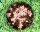
Mitochondria
21.11
Mitochondria are the cell's power producers. They convert energy into forms that are usable by the cell. They are the sites of cellular respiration which ultimately generates fuel for the cell's activities.

What are their distinguishing characteristics?
Mitochondria are bounded by a double membrane. Each of these membranes is a phospholipid bilayer with embedded proteins. The outermost membrane is smooth while the inner membrane has many folds. These folds are called cristae. The folds enhance the "productivity" of cellular respiration by increasing the available surface area.

Muscle Cell Mitochondria, Copyright Dennis Kunkel.
The double membranes divide the mitochondrion into two distinct parts: the intermembrane space and the mitochondrial matrix. The intermembrane space is the narrow part between the two membranes while the mitochondrial matrix is the part enclosed by the innermost membrane. Several of the steps in cellular respiration occur in the matrix due to its high concentration of enzymes.

Mitochondrion with matrix, Image courtesy of The Virtual Cell.
Mitochondria are semiautonomous (semi- auto-) in that they can divide and grow to make more of themselves. They also have their own DNA and ribosomes.
Share your opinions
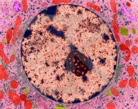
The Nucleus
21.03
 The nucleus is a membrane bound structure that contains the cell's hereditary information and controls the cell's growth and reproduction.
It is commonly the most prominent organelle in the cell.
Distinguishing Characteristics:
The nucleus is bounded by a double membrane called the nuclear envelope. This membrane separates the contents of the nucleus from the cytoplasm.The envelope helps to maintain the shape of the nucleus and assists in regulating the flow of molecules into and out of the nucleus through nuclear pores.
Chromosomes are also located in the nucleus.
 When a cell is "resting" i.e. not dividing, the chromosomes are organized into long entangled structures called chromatin and not into individual chromosomes as we typically think of them.
The Nucleolus:
 The nucleus also contains the nucleolus which helps to synthesize ribosomes.
The nucleolus contains nucleolar organizers which are parts of chromosomes with the genes for ribosome synthesis on them. Copious amounts of RNA and proteins can be found in the nucleolus as well.
The nucleus controls the synthesis of proteins in the cytoplasm through the use of messenger RNA. Messenger RNA is produced in the nucleolus of the cell and travels to the cytoplasm through the pores of the nuclear envelope.
cilia and flagella
21.00
 In Journey into the Cell, we looked at the structure of the two major types of cells: prokaryotic and eukaryotic cells.
Now we turn our attention to the "movers" of a eukaryotic cell, cilia and flagella.
What are cilia and flagella?
If the protrusions are short and numerous they are termed cilia. If they are longer and less numerous (usually only one or two) they are termed flagella.
What are some distinguishing characteristics?
The pattern is so named because a ring of nine microtubule "doubles" has in its center two singular microtubules.
This organization allows the sliding of the microtubule doubles against one another to "bend" the cilia or flagella. This type of organization is found in most eukaryotic cilia and flagella.
Where can cilia and flagella be found?
Cilia can be found in areas such as the respiratory and female reproductive tracts.
The Cell-Cell Structure
20.53
Eukaryotic Cells and Prokaryotic Cells
There are two primary types of cells: eukaryotic cells and prokaryotic cells. Eukaryotic cells are called so because they have a true nucleus. The nucleus, which houses DNA, is contained within a membrane and separated from other cellular structures. Prokaryotic cells however have no true nucleus. DNA in a prokaryotic cell is not separated from the rest of the cell but coiled up in a region called the nucleoid.As organized in the Three Domain System, prokaryotes include archaeans and bacteria. Eukaryotes include animals, plants, fungi and protists. Typically, eukaryoitc cells are more complex and much larger than prokaryotic cells. On average, prokaryotic cells are about 10 times smaller in diameter than eukaryotic cells.
Eukaryotes grow and reproduce through a process called mitosis. In organisms that also reproduce sexually, the reproductive cells are produced by a type of cell division called meiosis. Most prokaryotes reproduce through a process called binary fission. During binary fission, the single DNA molecule replicates and the original cell is divided into two identical daughter cells.
Both eukaryotic and prokaryotic organisms get the energy they need to grow and maintain normal cellular function through cellular respiration. Cellular respiration has three main stages: glycolysis, the citric acid cycle, and electron transport. In eukaryotes, most cellular respiration reactions take place within the mitochondria. In prokaryotes, they occur in the cytoplasm and/or within the cell membrane.
The Cell-Cell Structure
There are also many distinctions between eukaryotic and prokaryotic cell structure. The following table compares the cell structures found in a typical prokaryotic cell to those found in a typical animal eukaryotic cell.Cell Structure Comparison
| Eukaryotic and Prokaryotic Cell Structure | |||
| Cell Structure | Prokaryotic Cell | Typical Animal Eukaryotic Cell | |
| Cell Wall | Yes | No | |
| Centrioles | No | Yes | |
| Chromosomes | One long DNA strand | Many | |
| Cilia or Flagella | Yes, simple | Yes, complex | |
| Endoplasmic Reticulum | No | Yes (some exceptions) | |
| Golgi Complex | No | Yes | |
| Lysosomes | No | Common | |
| Mitochondria | No | Yes | |
| Nucleus | No | Yes | |
| Peroxisomes | No | Common | |
| Cell Membrane | Yes | Yes | |
| Ribosomes | Yes | Yes | |
Osmosis
09.25

- If the salt water solution is hypertonic it would contain a higher concentration of solute and a lower concentration of water than the blood cells. Fluid would flow from the area of low solute concentration (the blood cells) to an area of high solute concentration (water solution). As a result the blood cells will shrink.
- If the salt water solution is isotonic it would contain the same concentration of solute as the blood cells. Fluid would flow equally between the blood cells and the water solution. As a result the blood cells will remain the same size.
- If the salt water solution is hypotonic it would contain a lower concentration of solute and a higher concentration of water than the blood cells. Fluid would flow from the area of low solute concentration (water solution) to an area of high solute concentration (the blood cells). As a result the blood cells will swell and even burst.
Facilitated Diffusion
09.18
 Facilitated diffusion is a type of passive transport that allows substances to cross membranes with the assistance of special transport proteins. Some molecules and ions such as glucose, sodium ions and chloride ions are unable to pass through the lipid bilayer of cell membranes.
Through the use of ion channel proteins and carrier proteins that are embedded in the cell membrane these substance can be transported into the cell. Ion channel proteins allow specific ions to pass through the protein channel. The ion channels are regulated by the cell and are either open or closed to control the passage of substances into the cell. Carrier proteins bind to specific molecules, change shape and then deposit the molecules across the membrane. Once the transaction is complete the proteins return to their original position.gh the use of ion channel proteins and carrier proteins that are embedded in the cell membrane these substance can be transported into the cell. Ion channel proteins allow specific i
Diffusion and Passive Transport
08.29

Diffusion
Passive Transport
As illustrated in the image above: "This cartoon illustrates passive diffusion. The dashed line is intended to indicate a membrane that is permeable to the molecules or ions illustrated as red dots. Initially all of the red dots are within the membrane. As time passes, there is net diffusion of the red dots out of the membrane, following their concentration gradient. When the concentration of red dots is the same inside and outside of the membrane the net diffusion ceases. However, the red dots still diffuse into and out of the membrane, but the rates of the inward and outward diffusion are the same resulting in a net diffusion of O."- Steven Berg
Although the process is spontaneous, the rate of diffusion for different substances is affected by membrane permeability. Since membranes are selectively permeable (only some substances can pass), different molecules will have different rates of diffusion. For instance, water diffuses freely across membranes, an obvious benefit for cells since water is crucial to many cellular processes. Some molecules however must be helped across the cell membrane through a process called facilitated diffusion.
Animal Cells
07.52
 Animal cells are eukaryotic cells, or cells with a membrane-bound nucleus. Unlike prokaryotic cells, DNA in animal cells is housed within the nucleus. In addition to having a nucleus, animal cells also contain other membrane-bound organelles, or tiny cellular structures, that carry out specific functions necessary for normal cellular operation. Organelles have a wide range of responsibilities that include everything from producing hormones and enzymes to providing energy for animal cells.
Animal Cells: Structures and Organelles
The following are examples of structures and organelles that can be found in typical animal cells:- Centrioles - organize the assembly of microtubules during cell division.
- Cytoplasm - gel-like substance within the cell.
- Endoplasmic Reticulum - extensive network of membranes composed of both regions with ribosomes (rough ER) and regions without ribosomes (smooth ER).
- Golgi Complex - responsible for manufacturing, storing and shipping certain cellular products.
- Lysosomes - sacs of enzymes that digest cellular macromolecules such as nucleic acids.
- Microtubules - hollow rods that function primarily to help support and shape the cell.
- Mitochondria - power producers and the sites of cellular respiration.
- Nucleus - membrane bound structure that contains the cell's hereditary information.
- Nucleolus - structure within the nucleus that helps in the synthesis of ribosomes.
- Nucleopore - tiny hole within the nuclear membrane that allows nucleic acids and proteins to move into and out of the nucleus.
- Ribosomes - consisting of RNA and proteins, ribosomes are responsible for protein assembly.
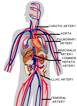
Anatomy of the Heart: Aorta
17.56
What is the aorta?
The aorta is the largest artery in the body. Arteries are vessels that carry blood away from the heart. The aorta arises from the left ventricle of the heart, forms an arch, then extends down to the abdomen, where it branches off into two smaller arteries.Function of the Aorta
The aorta carries and distributes oxygen rich blood to all arteries. Most major arteries branch off from the aorta, with the exception of the main pulmonary artery.Structure of the Aortic Walls
The walls of the aorta consist of three layers. They are the tunica adventitia, the tunica media, and the tunica intima.These layers are composed of connective tissue, as well as elastic fibers. These fibers allow the aorta to stretch to prevent over-expansion due to the pressure that is exerted on the walls by blood flow.
Branches of the Aorta
- Ascending Aorta - Extends from the left ventricle of the heart.
- Coronary Arteries
- Aortic Arch
- Brachiocephalic Artery
- Right Common Carotid Artery
- Right Subclavian Artery
Left Common Carotid Artery
Left Subclavian Artery - Right Common Carotid Artery
- Descending Aorta
Chest Region:
- Visceral Branches - Supply blood to the lungs, pericardium, lymph nodes, and esophagus.
- Parietal Branches - Supply blood to the chest muscles, diaphragm, and spinal cord.
Abdominal Region:
- Celiac Artery
- Left Gastric Artery - Supplies blood to the esophagus and portions of the stomach.
- Hepatic Artery - Supplies blood to the liver.
- Splenic Artery - Supplies blood to the stomach, spleen, and pancreas.
- Renal Arteries - Supplies blood to the kidneys.
- Ovarian Arteries - Supplies blood to the female reproductive organs.
- Testicular Arteries - Supplies blood to the male reproductive organs.
- Femoral Arteries - Supply blood to the lower legs and feet.
Superior Mesenteric Artery - Supplies blood to the intestines.
Inferior Mesenteric Artery - Supplies blood to the colon.
Common Iliac Artery - Visceral Branches - Supply blood to the lungs, pericardium, lymph nodes, and esophagus.
Capillary
08.55
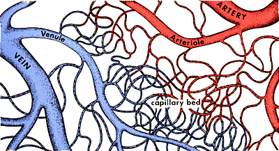
What Is a Capillary?
Capillary Size
Capillary Microcirculation
The flow of blood in the capillaries is controlled by structures called precapillary sphincters. These structures are located between arterioles and capillaries and contain muscle fibers that allow them to contract. When the sphincters are open, blood flows freely to the capillary beds of body tissue. When the sphincters are closed, blood is not allowed to flow through the capillary beds. Fluid exchange between the capillaries and the body tissues takes place at the capillary bed.
Ventricles of the Heart
08.53

Ventricles of the Heart
Function of the Ventricles
- Right ventricle: Receives blood from the right atrium and pumps it to the main pulmonary artery. The main pulmonary artery extends from the right ventricle and branches into left and right pulmonary arteries, which extend to the lungs. Here oxygen-poor blood picks up oxygen and is returned to the heart via the pulmonary veins.
- Left ventricle: Receives blood from the left atrium and pumps it to the aorta. The aorta carries and distributes oxygen-rich blood to the rest of the body.
Vein
08.47

A vein is an elastic blood vessel that transports blood from various regions of the body to the heart. Veins can be categorized into four main types: pulmonary, systemic, superficial, and deep veins.
Pulmonary veins carry oxygenated blood from the lungs to the left atrium of the heart. Systemic veins return deoxygenated blood from the rest of the body to the right atrium of the heart. Superficial veins are located close to the surface of the skin and are not located near a corresponding artery. Deep veins are located deep within muscle tissue and are typically located near a corresponding artery with the same name (for example coronary arteries and veins).
A vein can range in size from 1 millimeter to 1-1.5 centimeters in diameter. The smallest veins in the body are called venules. They receive blood from the arteries via the arterioles and capillaries. The venules branch into larger veins which eventually carry the blood to the largest veins in the body, the vena cava. The blood is then transported from the superior vena cava and inferior vena cava to the right atrium of the heart.
Artery
08.34
 An artery is an elastic blood vessel that transports blood away from the heart. There are two main types of arteries: pulmonary arteries and systemic arteries.
Pulmonary arteries carry blood from the heart to the lungs where the blood picks up oxygen. The oxygen rich blood is then returned to the heart via the pulmonary veins. Systemic arteries deliver blood to the rest of the body. The aorta is the main systemic artery and the largest artery of the body. It originates from the heart and branches out into smaller arteries which supply blood to the head region (brachiocephalic artery), the heart itself (coronary arteries), and the lower regions of the body.
The smallest arteries are called arterioles and they play a vital role in microcirculation. Microcirculation deals with the circulation of blood from arterioles to capillaries to venules (the smallest veins). The liver, spleen and bone marrow contain vessel structures called sinusoids instead of capillaries. In these structures blood flows from arterioles to sinusoids to venules.
Microcirculation
05.19

The precapillary sphincters contain muscle fibers that allow them to contract. When the sphincters are open, blood flows freely to the capillary bed where fluids, gasses, nutrients, and wastes are exchanged between the blood and body cells. When the sphincters are closed, blood is not allowed to flow through the capillary bed and must flow directly from the arteriole to the venule through the thoroughfare channel.
It is important to note that blood is supplied to all parts of the body at all times, but all capillary beds do not contain blood at all times. Blood is diverted to the parts of the body that need it most at a particular time. For instance, when you eat a meal blood is diverted from other parts of your body to the digestive tract to aid in digestion and nutrient absorption.
CERN on trial: could a lawsuit shut the LHC down?
05.16

COURTS and legal scholars love quoting legal maxims in Latin. One of the most famous is fiat justitia ruat caelum. The phrase is a resolute affirmation of the rule of law. It means "Let justice be done though the heavens fall".
It was intended as hyperbole. But, ironically, courts may now have to confront these words on literal terms. In various countries, plaintiffs have sought court orders to halt the operation of the Large Hadron Collider at CERN near Geneva, Switzerland, with the most extraordinary of allegations: that the experiment may create a black hole that will devour the Earth.
Up until now, the various lawsuits filed against the LHC have faltered. But if the right kind of claim is filed in the proper court, a judge may soon have to face the question of whether an injunction might be needed to save the world.
Injunctions are court orders that command persons to do or refrain from doing something. They are relatively routine, for example when a building of historic significance is threatened with demolition. But wading into the world of particle physics to shut down the LHC would be a forbidding proposition for anyone in judges' robes.
In deciding whether or not to issue an injunction, courts engage in what lawyers refer to as a "balancing test". The idea is that the court weighs the hardships that would be endured by both parties if the injunction were or were not issued, taking into account the likelihood and severity of the alleged consequences. The test closely resembles what is portrayed by courthouse statues around the world - Lady Justice holding up scales to measure the relative weight of the plaintiff's and defendant's cases.
So let's do the balancing test for the LHC case. The hardship CERN would suffer from an injunction is enormous - idling thousands of workers and equipment worth billions of euros, and upending a great scientific adventure. That weighs on the scales heavily. But on the other side is an Earth-mass black hole. That not only tips the scales, it eats them up.
The remaining task is to determine whether the questions raised are sufficiently serious. For that, a court must take a careful look at the scientific controversy. Yet the physics involved is difficult terrain even for physicists. A judge with maybe just a few days to ponder has scant chance of learning the science well enough to confidently decide who is right and who is wrong.
Usually when complex scientific issues are involved, courts turn to expert witnesses. But there is a problem with using experts in this case: none would seem to be without bias. CERN employs half of the world's particle physicists; the other half are their friends. All of them are anxiously awaiting data from the LHC to advance their field. The LHC is not just a particle physics experiment, it is the particle physics experiment. So what is a court to do?
Courts can maintain the rule of law in a fair and principled way by looking at the human context surrounding the scientific debate. While the physics may be largely impenetrable to the court, the human factors are not.
One question a court can investigate is how likely it is that the theoretical underpinnings of the scientific work are defective. Those seeking an injunction could, for example, ask a court to consider the history of shifting arguments for why the LHC is safe.
In 1999, physicists said no particle accelerator for the foreseeable future would have the power to create a black hole. But theoretical work published in 2001 showed that if hidden extra dimensions in space-time did exist, the LHC might create black holes after all. Thereafter, the argument for safety was changed. In 2003, it said that any black holes created would instantly evaporate. But when subsequent theoretical work suggested otherwise, the argument changed again. In 2008, CERN issued a report arguing a safety case based, ultimately, on astrophysical arguments and observations of eight white dwarf stars. These flip-flops on safety might cause a court to find current assurances less persuasive than they would otherwise be.
In addition, a court could look at the sociological and psychological context in which the disputed scientific work was carried out. Social scientists have identified a number of phenomena that can skew attempts to reach objective assessments of risk. For instance, cognitive dissonance describes the tendency of people to seek information that is consistent with their beliefs and to avoid information that is inconsistent. "Groupthink" describes a process by which intelligent individuals, working in a group, can reach a worry-free outlook that is not justified by the facts. And the phenomenon of confirmation bias - the tendency to filter information so as to confirm working hypotheses - was cited by the Columbia Accident Investigation Board as one explanation for why space shuttle programme managers ignored sure signs of trouble.
A court charged with deciding whether an injunction should be issued could consider whether these sorts of social effects plausibly undermine the conclusion that the LHC is safe.
These lines of inquiry might strike physicists as unfair. Many will argue that scientific work should be debated on its scientific merits alone. That objection is well put in a purely academic dispute, but the question of whether the LHC is safe is not academic - it is a real-world question with the highest possible stakes. Evaluating the science from a real-world perspective, and understanding scientific work to be a fallible human enterprise, is not merely fair - where justice is concerned, it is essential.
Mathematicians offer tip-offs to LAPD
05.14
 Los Angeles police are getting tip-offs from unlikely informants: mathematicians.
Using crime data from southern California, Jeffrey Brantingham of the University of California, Los Angeles, and his colleagues set out to calculate how the movements of criminals and victims create opportunities for crime, and how police can reduce it. They came up with a pair of equations that could explain how local crime hotspots form – which turned out to be similar to those that describe molecular reactions and diffusion.
The equations suggested that there are two kinds of hotspot. The first, called "supercritical", arises when small spikes in crime pass a certain threshold and create a local crime wave. The second, "subcritical", happens when a particular factor – the presence of a drug den, for instance – causes a large spike in crime. The equations also indicated that rigorous policing could completely eliminate the subcritical hotspots, but would simply displace the supercritical variety.
The approach "presents a novel hypothesis of how hotspots form", says John Eck, a criminologist at the University of Cincinnati, Ohio. Brantingham hopes eventually to be able to predict where subcritical hotspots are forming, so police can step in to nip problems in the bud. His team is already collaborating with Los Angeles police.
World's most sensitive neutrino experiment begins
05.10
 An intrepid subatomic particle has travelled through the bedrock of Japan and triggered a detector on the other side of the country, heralding a new attempt to probe the mystery of neutrino oscillations. The result could take us closer to answering one very big question – why is the universe full of matter?
In the "T2K" (Tokai-to-Kamioka) experiment, an intense beam of neutrinos is being generated in a particle accelerator near Tokai village north of Tokyo, and aimed at the Super-Kamiokande neutrino detector 300 kilometres away.
Neutrinos interact only reluctantly with matter, but from time to time one of the trillions in the beam will be lucky enough to hit an atomic nucleus inside Super-Kamiokande, and so create a distinctive flash of light.
The goal is to understand a strange kind of subatomic metamorphosis. These particles come in three types or flavours: electron, muon and tau neutrinos. From earlier experiments, physicists know that neutrinos spontaneously change their flavour, oscillating back and forth from one kind to another. But the details are still hazy.
Extremely sensitive
With T2K they are hoping to fill in some of the blanks. "We will be able to look for the appearance of electron neutrinos in a muon neutrino beam much more sensitively than has been done before," says David Wark of Imperial College, London. "It is ten times as sensitive as previous experiments."
This could shed some light on why we exist. "The known laws of physics should lead to roughly the same quantity of matter and antimatter in the universe," says Wark.
But because matter and antimatter destroy one another, that would have led to a cosmos full of radiation and almost no solid stuff. Instead matter somehow predominated. "That tells us there must be a law of physics that is different for matter and antimatter," Wark told New Scientist. "We don't know what it is, but neutrino oscillations are someplace where it might show up."
To test the idea, researchers will first measure what fraction of muon neutrinos in the T2K beam turn into electron neutrinos. Then T2K and other experiments could compare neutrinos and antineutrinos, to finally find out why the cosmos is so biased against antimatter.
If you would like to reuse any content from New Scientist, either in print or online, please contact the syndication department first for permission. New Scientist does not own rights to photos, but there are a variety of licensing options available for use of articles and graphics we own the copyright to.
Awas, Vila di Puncak Akan Dipangkas
04.49

Menteri Negara Lingkungan Hidup Gusti Muhammad Hatta menargetkan dapat menurunkan jumlah vila yang berada di kawasan puncak, Bogor, Jawa Barat.
"Kita harapkan setiap tahun ada penurunan jumlah vila," kata MenLH pada acara penanaman pohon di area Situ Cibeureum, Bojong Gede, Bogor, Jawa Barat, Jumat (8/1/2010).
Dia mengatakan, pihaknya membantu Pemkab Bogor sebagai ujung tombak pemilik daerah puncak untuk menata kembali kawasan itu sebagai daerah resapan air.
"Kita mencoba melakukan berbagai pendekatan, termasuk pendekatan persuasif. Kita tidak ingin mempermalukan orang," katanya.
MenLH tidak menyebutkan jumlah vila yang sekarang ada dan jumlah vila yang akan ditertibkan di daerah itu.
Sebelumnya, Satuan Polisi Pamong Praja (Satpol PP) mulai Selasa secara bertahap membongkar 87 vila liar di kawasan Cisarua, Kabupaten Bogor, Jawa Barat.
Pada kesempatan tersebut, MenLH beserta jajaran eselon I KLH dan Bupati Bogor Rahmat Yasin menyerahkan secara simbolis 250 bibit pohon trembesi, mahoni dan belimbing, serta 3000 ikan nila dan mujair di Situ Cibeureum.
Ada sekitar 850 bibit pohon yang bakal ditanam di kawasan Situ Cibeureum yang berasal dari sumbangan Yayasan Kehati, Solidaritas Istri Kabinet Indonesia Bersatu-2 dan KLH, yang selanjutnya akan dipelihara oleh masyarakat sekitar.
Harimau Sumatera Akan Dilepaskan Lagi di Hutan Tambling
04.41

Pelepasliaran harimau sumatera (Panthera tigris sumatrae) akan kembali dilakukan di kawasan Tambling Wildlife Nature Conservation (TWNC). Hutan konservasi yang terletak di Lampung bagian timur ini merupakan salah satu bagian dari hutan alam yang masih asli.
"Kami berencana melakukan pelepasliaran harimau dengan persiapan akhir pada 21 Januari 2010 dan dilepasliarkan pada 22 Januari 2010," kata Peggy Melati Sukma, juru bicara Artha Graha Peduli, dalam e-mail yang dikirim kepada Kompas.com, Rabu (13/1/2010). Saat ini, pengelolaan TWNC memang didukung lembaga sosial di bawah Artha Graha Group bekerja sama dengan Direktorat Jenderal Perlindungan Hutan dan Konservasi Alam Departemen Kehutanan.
Harimau-harimau yang dilepaskan di TWNC berasal dari Aceh dan Jambi. Ada enam harimau yang telah ditangkap di daerah konflik dengan penduduk di kedua daerah untuk dipindahkan ke habitat yang lebih sesuai. Dua ekor di antaranya, diberi nama Pangeran dan Agam, telah dilepaskan ke Hutan Tambling pada Juli 2008. Empat harimau lainnya, yang diberi nama Buyung, Panti, Ucok, dan Salma, akan dilepaskan menyusul pada waktu yang tepat.
TWNC menyediakan sarana untuk melatih harimau, termasuk enclosure field seluas 2,5 hektar, agar harimau tersebut tetap memiliki insting alami untuk dipastikan bertahan mandiri di hutan. Tim Taman Safari Idnonesia juga dilibatkan untuk memantau kesehatan harimau-harimau tersebut.
Tidak hanya harimau, kawasan TNWC seluas 45.000 hektar, yang merupakan bagian dari Taman Nasional Bukit Barisan Selatan, juga menyimpan spesies langka lain dan pemandangan yang alami. Hutan yang ada di kawasan tersebut dikategorikan sebagai hutan hujan tropika dataran rendah yang terdiri dari hutan pantai, hutan mangrove, dan rawa gambut. Sebagai besar kawasan itu merupakan zona inti dan cagar alam yang dijaga kelestariannya.
Hutan primer dan sekundernya menjadi habitat sekitar 50 jenis burung dari 11 familia. Sementara hutan mangrovenya menjadi tempat hidup tak kurang dari 41 spesies burung. Kawasan ini juga menjadi lokasi yang mendukung untuk populasi mamalia. Bagian pantainya merupakan tempat hidup penyu hijau, penyu belimbing, dan penyu sisik.
Matinya "Emi" Si Cula Dua
04.34

- Sungguh malang nasib Emi. Seekor badak Sumatera (Dicerorhinus sumatrensis) betina yang telah 14 tahun tinggal dan berkembang biak di Kebun Binatang Cincinnati, Ohio, Amerika Serikat itu mati pada 5 September 2009 di usia 21 tahun karena sakit. Kematian Emi juga menjadi duka bagi para ilmuwan dan orang-orang yang merawatnya selama ini.
Apalagi, Emi termasuk bagian dari kesuksesan program penangkaran badak secara ex-situ. Sebelumnya Emi adalah salah satu indukan yang baik dan telah melahirkan tiga keturunan di penangkaran. Masing-masing anaknya diberi nama Andalas, Suci, dan Harapan. Andalasa adalah badak jantan muda yang kini berada di Taman Nasional Way Kambas untuk mendukung program penangkaran badak Sumatera.
Kematian Emi adalah kasus kematian pertama badak Sumatera yang tinggal di penangkaran. Hal itu disampaikan Kepala Lembaga Penelitian Lindner Center for Conservation and Research of Endangered Wildlife (CREW) Cincinnati Zoo, Terri L. Roth saat jumpa pers Global Management and Propagation Board (GMPB) Meeting di Hotel Santika, Bogor, Jumat (15/1/2010).
"Kematian Emi karena mengalami gagal hati, di mana terdapat penumpukan zat besi pada jaringan tubuhnya," ujar Terri. Penyakit yang diderita Emi diketahui adalah hemokromatosis yang sering ditemukan pada beberapa hewan liar di penangkaran Cincinnati termasuk pada manusia, dan badak Afrika.
Matinya Emi, semakin mengurangi jumlah badak Sumatera yang saat ini diperkirakan hanya tinggal 200 ekor di dunia. Diperkirakan pula, 60 persen dari populasi badak Sumatera telah hilang selama 20 tahun terakhir.
Untuk badak Sumatera yang tinggal di penangkaran seperti Emi, jumlahnya baru sembilan ekor. Ditargetkan, dalam sepuluh tahun ke depan, jumlah badak Sumatera dapat bertambah tujuh ekor.
"Secara global, targetnya 17 badak Sumatera totalnya. Terhitung dari 2010 ini. Kita punya sekarang sepuluh, tapi hanya enam yang bisa berkembang biak. Diharapkan dari enam itu nambah tujuh lagi," ujar Direktur Yayasan Badak Indonesia (YABI), sekaligus kepala GMPB, Widodo Ramono.
Oleh karena itulah, saat ini GMPB terus melakukan upaya penyelamatan badak Sumatera melalui program Global Sumatran Rhino Propagation Program yang melibatkan seluruh negara dan institusi yang memiliki pusat penangkaran badak Sumatera, serta sponsor lainnya.
Rosa Lebih Tertarik dengan Manusia
04.31

BOGOR, Ada-ada saja ulah Rosa, seekor badak betina yang tinggal di penangkaran Suaka Rhino Sumatera (SRS) Way Kambas, Lampung, ini lebih tertarik dengan manusia ketimbang badak jantan.
"Kayak anjing peliharaan deh Mbak, mintanya diajak main. Dia emang deket banget sama manusia, beda sama yang lain. Pas kita di tenda, dia suka masuk ke tenda," ujar Kurnia, dokter hewan yang mengawal evakuasi Rosa dari Bukit Barisan Selatan ke Way Kambas saat berbincang seusai jumpa pers di Hotel Santika, Bogor, Jumat (15/1/2010).
Kedekatan Rosa dengan manusia justru menjadi masalah bagi pengembangbiakannya. Ketika didekatkan dengan badak jantan, Rosa menghindar dan justru menghampiri perawatnya. "Lebih tertarik dengan keeper-nya, itu jadi penghambat," kata Kurnia.
Ketua Yayasan Badak Indonesia (Yabi) Widodo Ramono juga mengaku tak habis pikir melihat ulah Rosa. "Rosa... rosa..., mungkin akan dibuat inseminasi buatan untuk Rosa, kalau tingkah lakunya begitu," kata Widodo.
Mengapa Rosa bertingkah aneh demikian, para peneliti masih mencari jawabannya. "Kita belum tahu, kita nemuin dia di (Taman Nasional) Bukit Barisan Selatan, dia asli BBS, mungkin ada cerita di balik itu," imbuh Kurnia.
Kurnia menduga, mungkin saat kecil Rosa telah ditinggal induknya. Oleh karena itulah, Rosa belajar dari manusia dan tak takut dengan manusia layaknya badak normal yang hidup soliter atau menyendiri. Ia mengatakan, jika perilaku Rosa terus-terusan abnormal seperti itu, dia tak siap untuk dikawinkan.
SRS Way Kambas memiliki lima ekor badak sumatera dalam penangkaran. Di antara lima badak yang ditangkar di sana, terdapat Andalas, badak sumatera pertama yang lahir di penangkaran sepanjang abad ke-19.
Macan Tutul Jawa Tertangkap Kamera TNGHS
04.30

Dua ekor macan tutul jawa (Panthera padus melas) dewasa tertangkap kamera yang dipasang tim pemantau macan Balai Taman Nasional Gunung Salak Halimun (TNGSH) belum lama ini. Hasil cetak foto dari kamera tersebut, juga mengungkap jalur jelajah satwa langka tersebut dilintasi oleh manusia.
"Untuk mengetahui populasi dan penyebaran macan tutul jawa kami melakukan monitoring dengan memasang camera trap pada 17 Desember 2009 hingga 5 Januari 2010. Hasilnya antara lain, terfoto oleh kamera tersebut dua ekor macan tutul jawa dewasa," kata Kepala Balai TNGHS Bambang Supriyanto di Bogor, Sabtu (16/1).
Lokasi pemantauan macan adalah kawasan resort Gunung Kendeng dengan koordinat S 0676542, E 9253778 (1165 m dpl) dan S 0681061, E 9253908 (1011 dpl). Metode yang dipakai adalah pemasangan camera trap serta pengamatan langsung terhadap marking, footprint, faeces, dan idenfikasi jenis pakan macan. Juga dilakukan wawancara dengan masyarakat setempat untuk mendapatkan informasi tambahan tentang keberadaan macan.
Selain memastikan keberadaan dua ekor macan dewasa, hasil monitoring juga menunjukan ketersediaan pakan bagi macan juga cukup. Kamera pun merekam keberadaan babi hutan dengan delapan ekor anaknya dan musang.
Marking (antara lain berupa bekas cakaran) dan footprint (jejak tapak kaki) yang ditemukan di areal pemantauan, menunjukan kawasan itu memang kawasan jelajah dan teritorial macan tutul jawa. Dari kotorannya yang ditemukan, jelas sekali satwa itu memangsa babi, musang, dan primata.
Kawasan TNGHS memang habitat terbaik bagi macan tutul jawa. Satwa ini pemangsa puncak pada rantai makanan (top predator) di kawasan hutan TNGHS. "Mereka mengendalikan hama babi dan musang di hutan taman nasional, sehingga tidak menjadi hama bagi petani yang bertani atau berkebun di sekitar kawasan," jelas Bambang.
Konflik macan dengan masyarakat, ungkap Bambang, terjadi pada Maret 2006 di Cisoka, Kabupaten Lebak dimana macan memangsa domba petani yang berakhir dengan kematian macan. Yang terakhir, sekor macan mati akibat diburu di kawasan yang masuk wilayah administratif Desa Kutajaya, Kecamatan Cijeruk, Kabupaten Bogor pada tahun 2009.
Mengenai seorang manusia dengan tangan membawa peralatan hendak berburu burung, yang tertangkap dalam kamera, Bambang memastikan, orang itu masuk kawasan taman nasional secara ilegal. Dan dipastikan pula, setiap orang yang disizinkan masuk kawan juga dilarang berburu atau mengambil apapun dari dalam kawasan.
"Sayang sekali, kamera menangkap orang itu dari belakang, sehingga sulit mengidentifikasi orang tersebut. Kami akan meningkatkan patrol hutan di kawasan itu dan penyadaran pada masyarakat di sekitar kawasan," kata Bambang, yang kehilangan dua camera trap dari 10 unit camera trap yang dipasang tim monitor populasi macan.
Kawasan TNGHS yang terletak di Kabupaten Bogor, Sukabumi, dan Lebak seluas 113,357 H . Pemantau populasi macan dengan metoda pemantauan seperti di kawasan hutan Resort Gunung Kendeng itu akan dilakukan di hutan kawasan taman nasional di setiap wilayah kabupaten tersebut. Saat ini populasi macan tutul jawa dipekirakan antara 41-45 ekor.
Induknya Kabur, Anak Orangutan Dipelihara Universitas
04.28

Pusat Penelitian Hutan Tropis Universitas Mulawarman, Samarinda, menyelamatkan seekor anak orangutan (Pongo pygmaeus) yang ditemukan di sebuah perkebunan kelapa sawit. Orangutan yang berusia sekitar satu tahun tersebut sempat telantar karena tidak ada pihak yang bersedia menampung.
Setelah telantar selama lebih dari 10 hari, anak orangutan jantan itu akhirnya dititipkan di Kebun Raya Universitas Mulawarman, Samarinda, Sabtu (16/1/2010). Kepala Pusat Penelitian Hutan Tropis Universitas Mulawarman Chandra Dewana Boer mengatakan, anak orangutan tersebut awalnya ditemukan oleh pegawai PT Agro Urea Sakti pada 5 Januari lalu.
Ketika itu, anak orangutan terlihat bersama induknya sedang mencari makan di lahan perkebunan yang masuk wilayah Kecamatan Sebulu, Kabupaten Kutai Kartanegara. Keduanya lalu diusir dari lahan perkebunan karena dikhawatirkan akan merusak tanaman sawit. Namun, induk orangutan kabur, sedangkan anaknya tertinggal di lokasi perkebunan.
Anak orangutan itu lalu dibawa ke perusahaan. Pada 10 Januari, anak orangutan itu diserahkan ke hutan pendidikan Penelitian dan Pengembangan Kehutanan Kementerian Kehutanan di Sebulu. ”Salah seorang peneliti Jepang dari Sumitomo Forestry yang ada di sana, Rio Soda, merekomendasikan supaya orangutan itu diselamatkan, jangan dilepaskan lagi di sekitar perkebunan itu karena di sekitar perkebunan sudah tidak ada hutan lagi,” kata Boer.
Selain tak ada jaminan dapat bertahan hidup, orangutan juga bisa ditangkap lagi oleh warga. ”Saya lalu menghubungi Yayasan Penyelamatan Orang Utan Borneo (Borneo Orang Utan Survival Foundation), tetapi di sana sudah penuh sehingga tidak bisa menampung orangutan baru,” kata Boer.
Pusat Penelitian Hutan Tropis Universitas Mulawarman lalu menghubungi Balai Konservasi Sumber Daya Alam Kalimantan Timur untuk meminta masukan. ”Karena belum ada solusi, lalu saya minta izin BKSDA untuk mengambil orangutan itu dari hutan penelitian Dephut. Sementara ini orangutan akan dititipkan di Kebun Raya Universitas Mulawarman dengan menggunakan berita acara,” kata Boer.
Sebelum dititipkan di Kebun Raya Universitas Mulawarman, orangutan itu sempat berada di kantor Pusat Penelitian Hutan Tropis Samarinda selama dua hari.
Tumbuhan Menjalar Ganggu Habitat Badak dan Hewan Liar Lainnya
04.28
Ada beberapa kendala dari luar yang menghambat perkembangbiakan badak Sumatra. Salah satunya adalah serangan tumbuhan menjalar mantangan (Meremia peltata) yang merambah habitat badak dengan cepat.
Hal tersebut disampaikan Kepala Yayasan Badak Indonesia (YABI), Widodo Ramono usai jumpa pers di Hotel Santika, Bogor, Jumat (15/1/2010). Menurut Widodo, meluasnya mantangan akan mempersempit habitat badak, gajah, atau harimau.
"Contohnya di Bukit Barisan Selatan, itu mantangannya sudah sangat meluas. Mereka (hewan) nggak suka ada mantangan, karena ada mantangan badak nggak jadi cari makan di sana, gajah, harimau juga," katanya.
Dijelaskan pula oleh Widodo, tumbuhan mantangan mirip dengan tanaman ubi jalar berdaun bulat yang merambat dan mampu melumpuhkan sebuah pohon besar. "Ini pohon besar, bisa mati sama mantangan, kalau enggak kuat. Mantangan cepat sekali meluasnya, entah kenapa, suatu kejadian yang nggak biasa, tumbuhnya bukan di tempat biasa. Mungkin karena perubahan ikllim," ujar Widodo.
Dalam jumlah kecil, mantangan tidak mengganggu badak karena akarnya dapat dimakan badak. Namun menurut Widodo, dalam jumlah besar seperti kondisi saat ini mantangan dapat mengganggu. "Kalau dimakan dalam jumlah besar, bisa jadi racun," katanya. Tumbuhan mantangan pun sulit dimusnahkan.
"Di Bukit Barisan Selatan pernah kita coba hilangkan dengan traktor, eh malah makin banyak. Setahun kemudian dilihat makin banyak," ujar Widodo. Widodo juga menyampaikan, para ilmuan akan terus meneliti pola hidup mantangan hingga menemukan cara mengontrol pertumbuhannya.
"Lagi diadakan penelitian, bekerja sama dengan pusat lembaga perhutanan. Kita ambil sampelnya di pot-pot kecil, kita amati," tutur Widodo. Oleh karena itu, Widodo mengusulkan kepada pemerintah untuk segera memperbaiki habitat badak Sumatra.
Ekosistem Bukit Soeharto Kian Terancam
04.26

Bukit Soeharto adalah kawasan yang menjadi hamparan hutan tropis basah dataran rendah yang pada Pemerintahan Orde Baru diharapkan jadi etalase kondisi hutan di Indonesia.
Karena letaknya mudah dijangau, yakni berada pada poros Jalan Lintas Kalimantan di Kaltim yang menghubungkan Balikpapan-Samarinda.
Penamaan yang sesuai dengan nama Presiden RI kedua itu tidak lepas dari sejarah bahwa Soeharto pernah melakukan perjalanan darat dari Balikpapan ke Samarinda melintasi bukit tersebut.
Penguasa Orde Baru itu juga sangat memperhatikan pengelolaan lingkungan di kawasan itu. Perhatian Soeharto itu diimplementasikan dengan berbagai kebijakan.
Pada kepemimpinan Soeharto, mulai dari 1976 telah terbit berbagai kebijakan yang intinya melindungi kawasan itu, sampai terbit lagi kebijakan terakhir pada 1994.
Kebijakan 1994 ini mengenai perubahan fungsi Taman Wisata Alam Bukit Soeharto seluas 61.850 hektar menjadi Taman Hutan Raya melalui Surat Keputusan Menteri Kehutanan nomor 419/Menhut-II/2004 tanggal 19 Oktober 2004.
Kawasan konservasi yang sempat menjadi kebanggaan Pemerintahan Orde Baru itu pernah dikunjungi sejumlah tamu penting atau tamu-tamu negara Indonesia untuk melihat keberhasilan Soeharto dalam menjaga kelestarian lingkungan, mulai dari Dubes, kepala negara termasuk Ratu Beatrix dari Belanda bersama suaminya.
Kawasan itu memiliki arti strategis bagi bidang ekologis karena menjadi wadah sebaran beberapa jenis flora antara lain Meranti (Shorea spp), Keruing (Dipterocarpus sp), Mahang (Hypoleuca), Mengkungan (Gigantea), Hora (Ficus sp), Medang (Lauraceae), Kapur (Dryobalanops spp), Kayu tahan (Anisoptera costata), Nyatoh (Palaquium spp), Keranji (Dialium spp) dan Perupuk (Laphopetalum solenospermum).
Taman Hutan Raya Bukit Soeharto juga menjadi habitat beberapa jenis satwa langka, antara lain Orangutan (Pongo pygmaeus), Beruang madu (Helarctos malayanus), Macan Dahan (Neofelis nebulosa), Landak (Hystrix brachyura) dan Rusa sambar.
Musibah kebakaran hutan/lahan yang kini rutin menimpa Bukit Soeharto ditambah dengan hadirnya 52 perusahaan KP yang mengkavling kawasan itu sudah tentu sangat mengancam keseimbangan ekosistem bukit Suharto.
Ekosistem Bukit Soeharto Kian Terancam
04.26
Bukit Soeharto adalah kawasan yang menjadi hamparan hutan tropis basah dataran rendah yang pada Pemerintahan Orde Baru diharapkan jadi etalase kondisi hutan di Indonesia.
Karena letaknya mudah dijangau, yakni berada pada poros Jalan Lintas Kalimantan di Kaltim yang menghubungkan Balikpapan-Samarinda.
Penamaan yang sesuai dengan nama Presiden RI kedua itu tidak lepas dari sejarah bahwa Soeharto pernah melakukan perjalanan darat dari Balikpapan ke Samarinda melintasi bukit tersebut.
Penguasa Orde Baru itu juga sangat memperhatikan pengelolaan lingkungan di kawasan itu. Perhatian Soeharto itu diimplementasikan dengan berbagai kebijakan.
Pada kepemimpinan Soeharto, mulai dari 1976 telah terbit berbagai kebijakan yang intinya melindungi kawasan itu, sampai terbit lagi kebijakan terakhir pada 1994.
Kebijakan 1994 ini mengenai perubahan fungsi Taman Wisata Alam Bukit Soeharto seluas 61.850 hektar menjadi Taman Hutan Raya melalui Surat Keputusan Menteri Kehutanan nomor 419/Menhut-II/2004 tanggal 19 Oktober 2004.
Kawasan konservasi yang sempat menjadi kebanggaan Pemerintahan Orde Baru itu pernah dikunjungi sejumlah tamu penting atau tamu-tamu negara Indonesia untuk melihat keberhasilan Soeharto dalam menjaga kelestarian lingkungan, mulai dari Dubes, kepala negara termasuk Ratu Beatrix dari Belanda bersama suaminya.
Kawasan itu memiliki arti strategis bagi bidang ekologis karena menjadi wadah sebaran beberapa jenis flora antara lain Meranti (Shorea spp), Keruing (Dipterocarpus sp), Mahang (Hypoleuca), Mengkungan (Gigantea), Hora (Ficus sp), Medang (Lauraceae), Kapur (Dryobalanops spp), Kayu tahan (Anisoptera costata), Nyatoh (Palaquium spp), Keranji (Dialium spp) dan Perupuk (Laphopetalum solenospermum).
Taman Hutan Raya Bukit Soeharto juga menjadi habitat beberapa jenis satwa langka, antara lain Orangutan (Pongo pygmaeus), Beruang madu (Helarctos malayanus), Macan Dahan (Neofelis nebulosa), Landak (Hystrix brachyura) dan Rusa sambar.
Musibah kebakaran hutan/lahan yang kini rutin menimpa Bukit Soeharto ditambah dengan hadirnya 52 perusahaan KP yang mengkavling kawasan itu sudah tentu sangat mengancam keseimbangan ekosistem bukit Suharto.
Danau Sentani Papua Septictank Besar Warga
04.25

Alam kebanggaan Papua, Danau Sentani di Jayapura perlahan namun pasti sedang menuju kerusakannya. Danau dengan pulau-pulau kecil di dalamnya ini terancam oleh sedimentasi/pendangkalan akibat aktivitas di Pegunungan Cagar Alam Cycloops, sampah rumah tangga, hingga sampah bahan beracun berbahaya.
"Danau Sentani ini juga sudah mirip dengan septictank besar, tempat pembuangan kotoran manusia yang tinggal di tepi-tepi danau," ujar Franz Albert Yoku, Ketua Umum Badan Otorita Adat Sentani (BOAS), Selasa (19/1/2010) di Sentani Kabupaten Jayapura Papua.
Ini diungkapkannya dalam peresmian BOAS yang dilakukan Gubernur Papua Barnabas Suebu. Franz menuturkan, kondisi Danau Sentani semakin terancam oleh pertambahan penduduk yang tidak memiliki keterampilan. Ini membuat warga cenderung mengambil cara gampang untuk mengeksploitasi alam yang merusak.
Karenanya, Franz Albert menargetkan agar penduduk Sentani memperoleh pendidikan serta keahlian untuk mengelola dan menjaga alamnya. Lebih lanjut, ia pun berusaha agar Pegunungan Cycloops bebas dari permukiman penduduk, ladang/kebun, dan berbagai aktivitas manusia.
Aktivitas di Cycloops menyebabkan tanah tergerus sehingga turun ke sungai dan terbawa ke Danau Sentani. Kondisi yang berlangsung terus menerus ini dikhawatirkan menyebabkan pendangkalan di danau. Sementara itu, Gubernur Papua Barnabas "Bas" Suebu meminta agar BOAS menjaga adat istiadat serta budaya dan kearifan lokal masyarakat Sentani.
Bas yang juga asli dari suku di Sentani mengatakan nilai-nilai moral, sosial, serta etika bermasyarakat dalam masyarakat adat harus diperhatikan. Ia mencontohkan, kini banyak anak-anak suku di Sentani yang tak bisa lagi berbicara dalam bahasa lokal suku.
"Ini yang dapat menjadi kepunahan," ujar Bas. Ihwal kerusakan alam Hutan Lindung Cycloop, Bas mengatakan Pemprov Papua telah menyiapkan strategi pembangunan kota baru yang menjauhi Cycloop yaitu ke arah selatan-barat Danau Sentani. Ini diharapkan dapat mengendalikan kegiatan ke arah Cycloop.
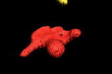
Sadis, Bunga Raflesia Ini Dipotong Satu Kelopaknya
04.23

Bunga Raflesia (Raflesia arnoldi) yang sedang mekar sempurna di Km 40 jalan lintas Kota Bengkulu-Kepahiang dirusak oknum tak bertanggungjawab dengan memotong salah satu kelopak bunganya.
Anggota tim Peduli Puspa Langka Tebat Monok Kabupaten Kepahiang, Holidin mengatakan bunga tersebut mekar hanya berjarak 2 meter dari badan jalan sehingga sangat mudah dijangkau pengunjung.
"Kami tidak tahu siapa yang memotong kelopak ini karena tidak ada penjagaan saat malam hari,"katanya, Senin.
Bunga langka yang mekar sempurna itu masih ramai didatangi pengunjung yang ingin melihat keindahan bunganya.
Salah seorang pengunjung, Musiardanis mengatakan merasa kecewa dengan tindakan tidak terpuji itu.
"Bangsa lain ada yang mengklaim bunga ini saking unik dan langkanya, tetapi masyarakat kita sendiri sama sekali tidak memiliki rasa tanggung jawab untuk memelihara, malah merusak,"katanya.
Bunga Raflesia yang normalnya memiliki lima kelopak, dengan dipotongnya satu kelopak tersebut hanya tinggal empat sehingga membuat bunga itu kurang menarik untuk dipandang.
Menurut Kabag Tata Usaha Balai Konservasi Sumber Daya Alam (BKSDA) Provinsi Bengkulu, Supartono mengatakan Hutan Lindung (HL) Bukit Daun merupakan habitat Bunga Raflesia dan memang sering ditemui bunga yang sedang mekar.